

ShapeLogic v 1.2 contains a generic particle analyzer . This page illustrates a few examples of the types of images that can currently be handled, and the types of images that the next versions of ShapeLogic should be able to handle.
All images are from
If you have a suggestion about what types of cell images would be important to handle next you can leave a message on the ShapeLogic mailing list .
Sparse cell images without categorizer
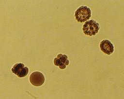
embryos.jpg a sample image from ImageJ. The un-tweaked particle counter or analyzer in ShapeLogic 1.2 worked with the default parameter values:
_iterations = 2; // K-mean iterations for color hypothesis
_maxDistance = 50; // Distance between color in same color bucket
_minPixelsInArea = 30; // Minimum number of pixels in particle
_maxPixelsInArea = 10000; // Maximum number of pixels in particle
Does not separate overlapping embryos or categorize them.
The particle counter or analyzer find 85 particles, which is consistent with what you get by counting by hand.
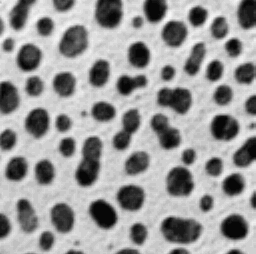
blobs.gif a sample image from ImageJ works with the default parameters.
Does not separate overlapping blobs or categorize them.
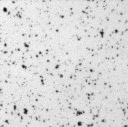
Cell_Colony.jpg a sample image from ImageJ works with the default parameters.
It is hard for a human to count the particles in this image but ShapeLogic v 1.2 gave a reasonable result.
Does not separate overlapping cells or categorize them.
Sparse cell images with categorizer. These images are handled by the current particle analyzer by adding rules to categorize. This can be done by the users, but a few applications might be added.
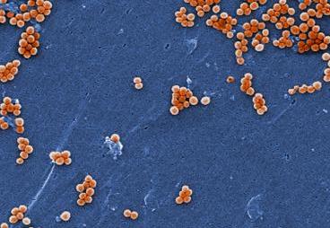
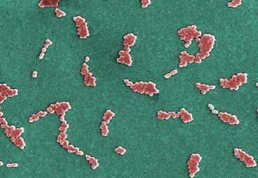
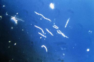
A low hanging fruit is a bacteria categorizer that can separate spherical bacteria, called cocci or rod-shaped, called bacilli.
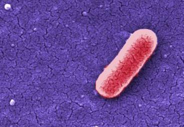
CDC image 10067
This should be categorized as a bacillus or a rod bacteria.
Making these rule should be simple, the big problem is that bacteria tend to lump together.
In the particle analyzer a particle's outline is found and turned into a polygon. While some cell parts or whole cell would be better described as a line.
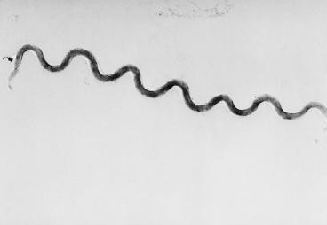
CDC image 2333
If it turns out to be too hard to handle them as an outline ShapeLogic might treat them as a line.
Bacteria, both cocci and bacilli, will often lump together. This make the problem of handling them a lot harder. Initially this will have to be handled by specialized particle analyzers.
Some medical images have a strong background gradient. There are standard techniques for handling this problem, they have not been combined with the ShapeLogic particle analyzer yet.
CDC image 6631
This demands that the particle analyzer understand what is inside and what is outside a given cell or virus.
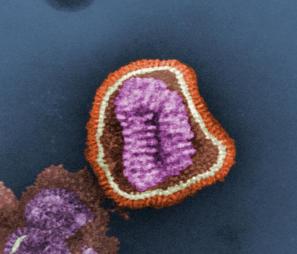
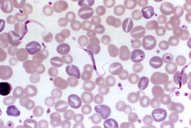
CDC image 10073
A influenza virus particle.